Fig. 2.
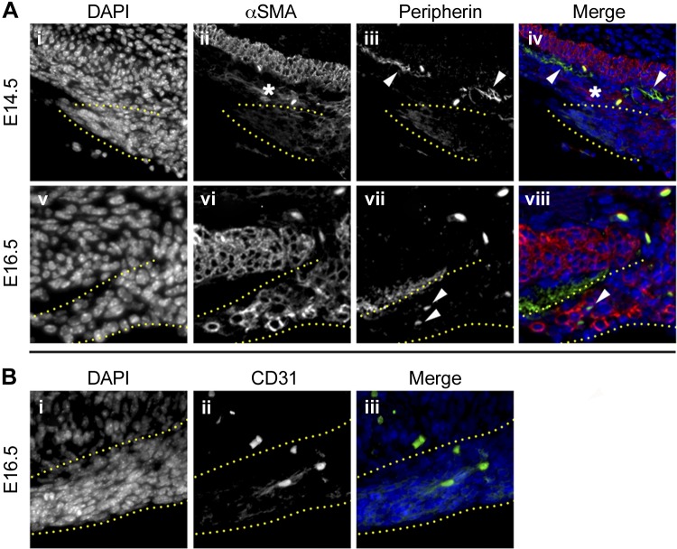
The gastric ligaments are predominantly smooth muscle structures. Immunofluorescence of WT pylorus at E14.5 (Ai–iv) and 16.5 (Av–viii, Bi–iii). Stains are listed above images. Ai–iv: at E14.5, α-smooth muscle actin (α-SMA) expression is low or absent within the developing gastric ligaments (outlined by yellow dots) and in the region of the gastric outer longitudinal muscle (OLM) (asterisk, Aii), since the smooth muscle has not yet matured. Comparatively, the mature gastric inner circular longitudinal muscle (ICM) strongly expresses α-SMA at this time point (bright staining above asterisk in Aii; red in Aiv). Peripherin expression is largely limited to the myenteric plexus adjacent to the gastric ICM (arrowheads in Aiii–iv). Av–viii and Bi–iii: at E16.5, the intensity of α-SMA staining in the gastric ligaments (outlined by yellow dotted lines) has increased, indicating robust smooth muscle differentiation (Avi and viii). However, there is limited expression of peripherin (arrowheads in Avii and Aviii) or CD31 (Bii and Biii), indicating that scant neurovascular tissue is present in the developing gastric ligaments. Orientation is as follows: stomach is left, and dorsal is up.

