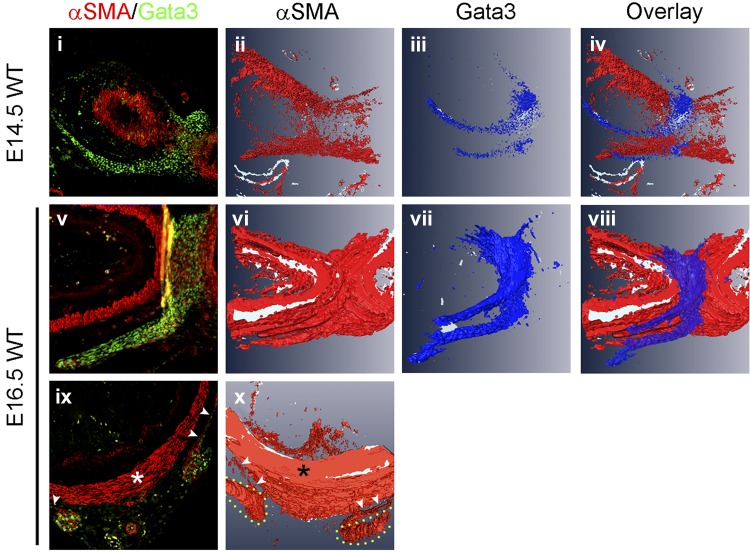
Fig. 4.
The gastric ligaments are physically contiguous with the pyloric OLM. Individual sections (i, v, and ix) and 3-dimensional (3-D) image reconstruction (ii–iv, vi–viii, and x) of WT pylorus based on α-SMA and GATA3 immunofluorescence. i and iv: Superficial longitudinal sections at E14.5 (i) and E16.5 (iv) stained for α-SMA (red) and GATA3 (green). GATA3 expression at both time points highlights the physical contiguity between the gastric ligaments and pyloric OLM. ii–iv and vi–viii: At E14.5, 3-D image reconstruction emphasizes the absence of α-SMA expression in the gastric ligaments and shows that GATA3 is expressed in a saddle-shaped domain at the pylorus, the ends of which comprise the gastric ligaments. Two days later at E16.5, the smooth muscle of the OLM and gastric ligaments has matured. The GATA3 expression pattern has slightly expanded but is otherwise unchanged. At this time point, the α-SMA and GATA3 expression domains essentially completely overlap within the gastric ligaments. ix: Cross section at E16.5, stained for α-SMA (red) and GATA3 (green). The thick, α-SMA-positive pyloric ICM completely surrounds the pylorus (asterisk). The pyloric OLM (small white arrowheads) is a thin layer laterally and absent ventrally. GATA3 staining (green) is visible in the thin lateral OLM (small white arrowheads) and in the contiguous round structures that represent cross sections of the gastric ligaments. Physical continuity is visible between the pyloric OLM and the gastric ligaments (particularly on the right side of this section). x: 3-D reconstruction of α-SMA-stained regions emphasizes the continuity between the thin pyloric OLM (small white arrowheads) and the pyloric ligaments (outlined in yellow), with virtually absent ventral OLM. The pyloric ICM is a distinct muscular structure (asterisk).

