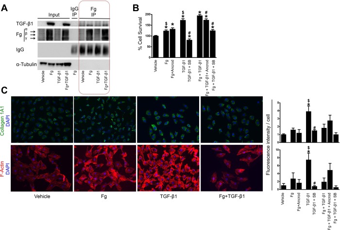
Fig. 4.
Fg treatment leads to transforming growth factor (TGF)-β1 expression and proliferation of cultured fibroblasts. A: immunoprecipation with anti-Fg antibody in NRK-49F cells after treatment with Fg, TGF- β1, or the combination of both. B: cell survival as evaluated by MTT assay in NRK-49F cells after treatment with Fg, TGF-β1, or the combination of both. The effects of additional Fg inhibition (Ancrod) and TGF-β1 inhibition [SB-431542 (SB)] on these treatments were also evaluated. n = 12 replicates/group (6 replicates for each condition from 2 experiments). *P < 0.05 compared with vehicle; #P < 0.05 compared with treatment without inhibitor; $P < 0.05 compared with Fg + TGF-β1 treatment. C: representative immunostaining of Col1A1- and phalloidin-based staining of F-actin in NRK-49F cells after treatment with Fg, TGF-β1, or the combination of both. DAPI, 4′,6-diamidino-2-phenylindole. Fluorescence quantification results are shown for these as well as combination treatments with Fg and TGF-β1 inhibitors (data represent 2 independent experiments with 2 replicates each for a total of 4 replicates; 5 images/replicate were taken, and the results were averaged). Data are presented as means ± SE of fold changes from vehicle. *P < 0.05 compared with vehicle; #P < 0.05 compared with treatment without inhibitor; $P < 0.05 compared with Fg + TGF-β1 treatment.

