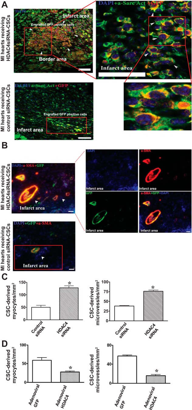
Fig. 8.
HDAC4 inhibition affects c-kit+ CSC-derived myocardial regeneration in infarcted hearts. A: the representative images of c-kit+ CSC-derived cardiomyocytes in MI hearts that received HDAC4 siRNA-treated and control siRNA-treated c-kit+ CSCs, respectively. c-kit+ CSCs were labeled with GFP, cardiomyocytes were stained with α-sarcomeric actinin (green), GFP was stained with anti-rabbit-IgG-Cy-3 (red), and nuclei were stained with DAPI (blue). B: the representative images of c-kit+ CSC-derived microvessels in MI hearts received HDAC4 siRNA-treated and control siRNA-treated c-kit+ CSCs, respectively. Microvessels were stained with α-smooth muscle actin (α-SMA) (red), GFP was stained with anti-rabbit-IgG-FITC (green), and nuclei were stained with DAPI (blue). C: quantitative analyses of c-kit+ CSC-derived myocytes and microvessels in MI hearts that received HDAC4 siRNA-treated and control siRNA-treated c-kit+ CSCs, respectively. D: quantitative analyses of c-kit+ CSC-derived myocytes and microvessels in MI hearts that received adenoviral HDAC4 and adenoviral GFP infected c-kit+ CSCs, respectively. The details of the immunostaining procedure are described in materials and methods. The number of c-kit+ CSC-derived myocytes and microvessels were counted in 5–6 randomized fields of 3 tissue sections for each heart. These sections, which contained infarct and border regions, were taken in the middle plane of each heart and were normalized to the tissue area. The values represent means ± SE (n = 3–5 per group). *P < 0.05 vs. MI heart that received control siRNA-treated c-kit+ CSCs in C. *P < 0.05 vs. MI hearts that received adenoviral GFP-infected c-kit+ CSCs in D. Scale bars represent 50 μm.

