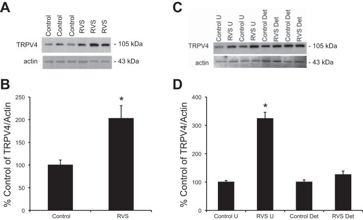
Fig. 2.
Increased TRPV4 expression in whole urinary bladders (A and B) and in urothelium (U; C and D) following 7 d of RVS as detected by Western blotting techniques. Representative Western blots (A and C) and summary histograms (B and D) of TRPV4 expression in whole urinary bladders (35 μg) and urothelium and detrusor smooth muscle from control rats and RVS-treated rats. Actin expression was used as a loading control. Relative TRPV4 band density in each group was normalized to actin expression and expressed as a percentage (%) of control in the same samples. RVS significantly (P ≤ 0.01) increased TRPV4 expression compared with control in whole urinary bladder and urothelial tissue but not detrusor smooth muscle. Statistical analyses were performed on raw data using Student's t-test (whole bladder) or ANOVA (split bladder, i.e., urothelium and detrusor) as described in Statistical Analyses. Values are means ± SE. Sample sizes are n of 7; *P ≤ 0.01.

