Fig. 5.
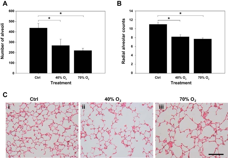
Quantification of numbers of alveoli and radial alveolar counts (RAC) in control and hyperoxia-exposed mice. Note: the numbers of alveoli (A) and RAC were (B) decreased in 40% oxygen and 70% oxygen-exposed mice compared with control mice. There was no difference in the number of alveoli and RAC between the lungs of 40% and 70% oxygen-exposed mice. Representative sections (C) of paraffin-embedded lungs from each treatment group stained with hematoxylin and eosin are also shown (i, ii, and iii represent control, 40%, and 70% oxygen, respectively; bar = 20 μm). *Significant difference, P < 0.05.

