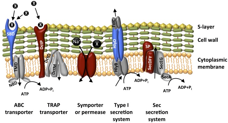
Figure 5.
Model of the localization of surface proteins involved in transport in D. reducens. Gray coloring indicates proteins expected to belong to the transport complex, but that were not identified in our extracts, while colored proteins are those identified: blue coloring indicates lipoproteins, red indicates membrane spanning proteins; black circles represent solutes (unspecified, if marked with an X). SBP, solute binding protein; NBD, nucleotide binding domain; SP, signal peptidase.

