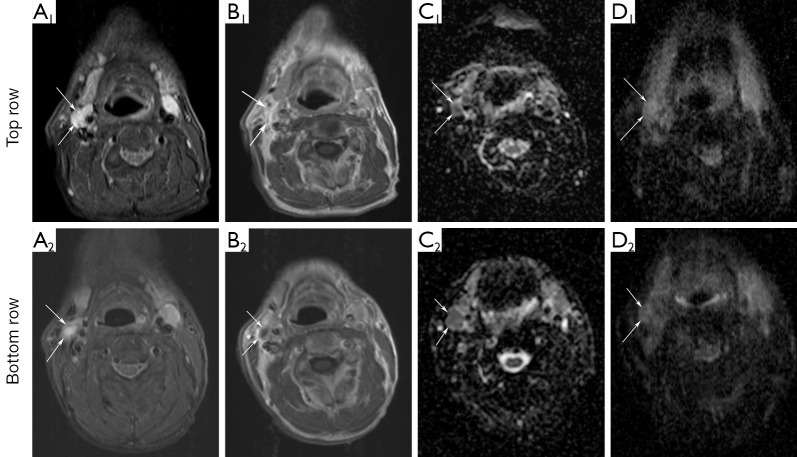
Figure 3.
Axial images showing a metastatic node (arrows) in patient number 1 in whom recurrent viable squamous cell carcinoma was diagnosed histopathologically in level II right during follow-up. DW-MRI1 (top row) and DW-MRI2 (bottom row): (A) STIR; (B) contrast-enhanced T1WI; (C) ADC maps with EPI technique and (D) ADC maps with HASTE technique. ADCEPI-values of the lymph node (arrow) are 99×10–5 and 102×10–5 mm2/s for DW-MRI1 and DW-MRI2, respectively. ADCHASTE-values are 106×10–5 and 118×10–5 mm2/s. Four years after completion of CRT this patient died due to lung metastases.

