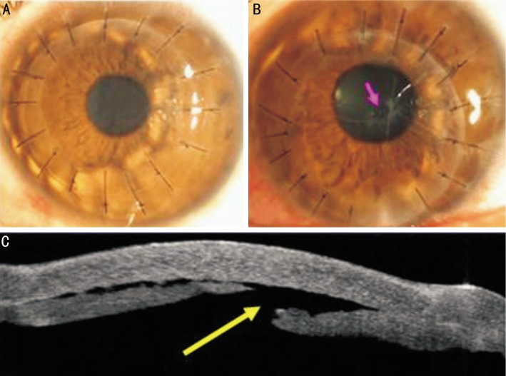
Figure 4. Slit-lamp photograph of FDALK for keratoconus showed a clear graft at postoperative day 5.
A:Slit-lamp photograph of small perforation of Descemet's membrane (red arrow) at postoperative 4.5wk; B: Anterior segment optical coherence tomography of small perforation of Descemet's membrane (yellow arrow) at postoperative 2wk.

