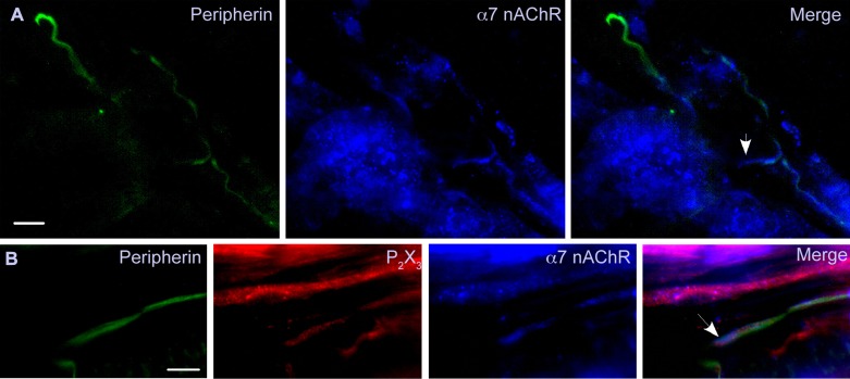
Fig. 7.
Group IV fibers within the muscle express α7-nAChR. Sections are shown from 2 gastrocnemius muscles (different animals) that were labeled with antibodies directed against peripherin (green), α7-nAChR (blue), and P2X3R (red). A: several peripherin-positive fibers (group IV) are shown, but only those in the lower right corner were also positive for α7-nAChR. In the merged image (right), the arrow shows α7-nAChR labeling extending beyond that of peripherin into an apparent axon terminal region. B: a peripherin-positive fiber is also stained with the α7-nAChR and P2X3R antibodies. The α7-nAChR and P2X3R labeling appears to extend beyond that of peripherin to the terminal end of the fiber (marked by arrow). There appears to be a parallel fiber that is only positive for P2X3R, which could be a group III fiber since it was not positive for peripherin. The bar in the lower corner of each peripherin panel indicates 12.5 μm. The examples shown here are representative of labeling experiments done on gastrocnemius muscle tissue from the 3 animals for both α7-nAChR and P2X3R.

