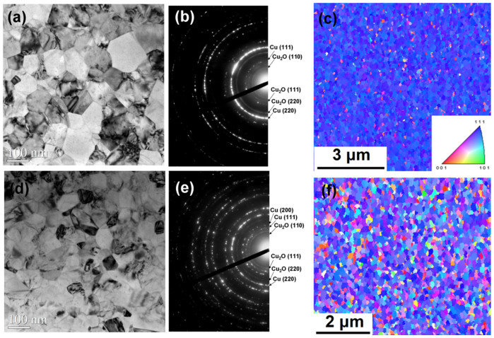
Figure 1.
Microstructure analysis for the two Cu seed layers in this study: (a) Plan-view TEM image for the strong <111>-oriented seed layer. (b) The corresponding SAD of the grains in (a). (c) Plan-view EBSD orientation image map of the strong <111>-oriented seed layer. (d) Plan-view TEM image for the regular <111>-oriented seed layer. (e) The corresponding SAD of the grains in (d). (f) Plan-view EBSD orientation image map of the regular <111>-oriented seed layer.

