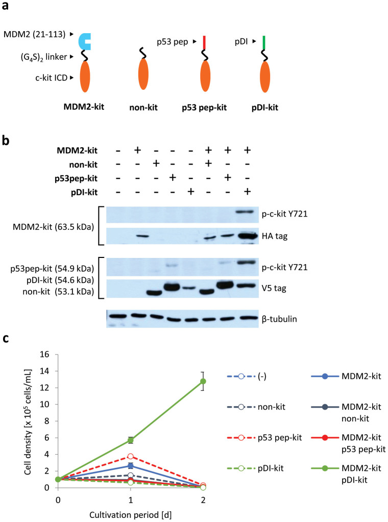
Figure 4. Detection of the interaction between MDM2 and its peptide inhibitor.
(a) Illustration for the constructed chimeras. The N-terminal domain of MDM2 and its peptide inhibitors (p53 pep and pDI) were fused to c-kit ICD. (b) Tyrosine phosphorylation of c-kit ICD of chimeras. Western blot analysis was performed with anti-phospho Y721 of c-kit to detect the phosphorylated form of the chimeras, and with an anti-HA or an anti-V5 tag to detect the expression level of the MDM2-fused chimeras (HA tag) or the others (V5 tag). The blots for β-tubulin are indicated as a loading control. (c) Growth of the cells expressing chimeras. Initial cell density was 1 × 10⁵ cells/mL. The viable cell densities are indicated as mean ± SD (n = 3, biological replicates).

