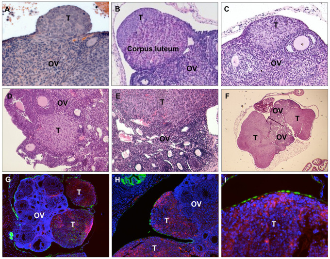
Figure 3. Intrauterine injection of OTICs forms in situ ovarian cancer in superovulated mice.
(A–F). H&E staining of mouse ovaries with tumors formed by OTICs. (G–I). Immunofluorescence staining of mouse ovaries with mCherry tumors. (Green, CK8. Red, mCherry. Blue, DAPI. OV, ovary. FT, fallopian tubes. T, tumor.)

