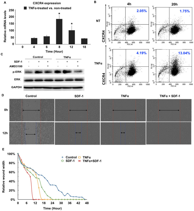
Figure 5. TNFα enhances the SDF-1/CXCR4 signaling in OTICs.
(A). Quantitive RT-PCR of CXCR4 in OTICs treated with TNFα. *P<0.05 vs control. (B). Flowcytometry analysis of cell surface CXCR4. Non-treated (NT) and TNFα-treated (TNFα) OTICs were stained with Alexa Fluor 594-labeld anti-CXCR4 antibody (FL2) and analyzed at 4 h and 20 h. The percentage of cells expressing high levels of CXCR4 was indicated in the graphs. (C). Western blots of p-ERK and total ERK in OTICs. GAPDH was used as loading control. The full-length blots are included in supplementary information. (D). Images of wound healing assay. (E). Wound width-time dependent curves for wound healing assay of OTICs.

