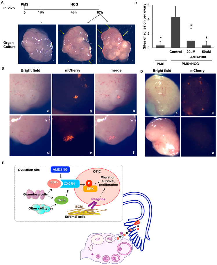
Figure 8. Ovulation is associated with ovarian tumor formation.
(A). Images of mouse ovaries. 19 hours after injecting PMS, the ovaries contain many mature follicles (a). 48 hours after injecting PMS, HCG was injected to trigger ovulation. 19 hours after injecting HCG, the ovaries contain many ovulating follicles (b, yellow arrows). (B). Images of mCherry-OTICs attaching to the ovulatory sites after 24-hour co-culture with mouse ovaries. (a, d, ovulatory sites; b, e, mCherry-OTICs; c, f, merged images.) (C), Numbers of the mCherry-OTIC adhesion sites on mouse ovaries. The numbers were counted as sites of adhesion per ovary. (PMS, ovaries 19 hours after PMS injection. PMS+HCG, 19 hours after HCG injection. Control, co-culture without AMD3100. AMD3100, co-culture with AMD3100 (20 uM or 50 uM) in the medium. n = 3. *P<0.05 vs PMS+HCG control group) (D). AMD3100 inhibited the adhesion of mCherry-OTICs to the ovary. (a, b, control; c, d, AMD3100-treated.) (E). Schematic model of the attraction and adhesion of extra-ovarian cancer cells to the ovary.

