
FIG. 2.
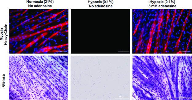
Representative images of myotubes formed only in hypoxia-survived C2C12 cells in the presence of adenosine. C2C12 cells underwent identical testing conditions, as described in Figure 1, except that these cells were cultured in the complete growth medium for 3 days after transfer to normoxic conditions followed by a 2% horse serum-containing differentiating medium for 6 days. C2C12 cells were immunostained (Upper) with MF-20 (myosin heavy chain, red) and DAPI (nuclei, blue), and stained with Giemsa (Lower) (scale bars=200 μm). Color images available online at www.liebertpub.com/tea