
Figure 2.
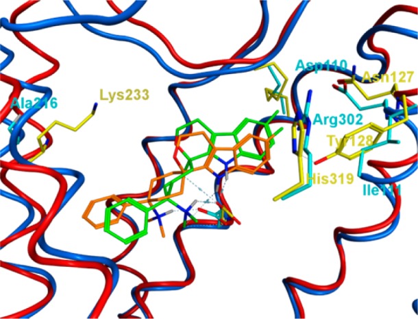
Three-dimensional structural overlay of the binding modes of 3a in the NOP and MOP receptors. NOP: Binding modes of 3a (orange) in the NOP receptor: NOP ribbons, blue; residues, cyan. MOP: Binding modes of 3a (green) in the MOP receptor: MOP ribbons, red; residues, yellow. MOP ribbons of helix 7, His319/Arg302, removed for clarity.