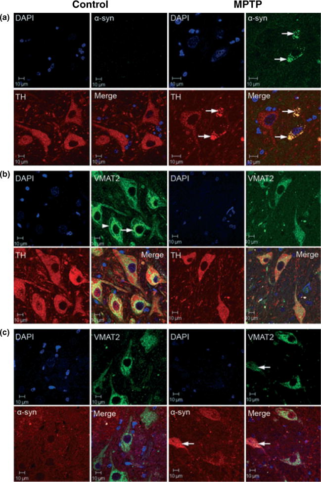
Fig. 6.

Confocal microscopy images of α-synuclein, TH and VMAT2 immunofluorescense labeling in control and MPTP-treated brains at the level of the substantia nigra pars compacta (coronal section) (Scale bar = 10 μm). (a) TH (red), α-synuclein (green), DAPI (blue) and merged triple labeling. There is a significant amount of α-synuclein punctate staining (or aggregation) co-localized with TH immunostaining in the cytosol of neurons from MPTP-treated animals that appear to be undergoing degeneration (see arrows). The insert in the α-synuclein panel from MPTP-treated animals is representative of the occasional α-synuclein-positive Lewy body-like spherical aggregates observed in MPTP-treated animals. (b) DAPI (blue), VMAT2 (green), TH (red) and merged triple labeling. In neurons from control animals, VMAT2 staining expressed a distinct subcellular pattern with a band labeling pattern in perinuclear regions (see arrow) and adjacent to the plasma membrane (see arrow head); the latter may be representative of a releasable pool of secretory vesicles. In nigral neurons from MPTP-treated animals, VMAT2 labeling expressed a more homogeneous distribution throughout the cytoplasm with disruption of the distinct labeling pattern observed in neurons from control animals. (c) DAPI (blue), VMAT2 (green), α-synuclein (red) and merged triple labeling. α-synuclein co-localized with VMAT2 as aggregates in the cytoplasm of degenerating neurons in MPTP-treated animals (see arrows).
