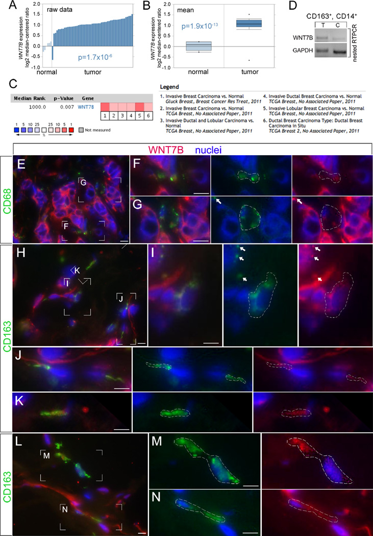
Fig 1. WNT7B expression in human mammary tumors.
WNT7B expression (as log2 median centered ratio) for mammary gland, normal and tumor stroma, shown either as raw data (A) or as the mean (B). In the chart showing the mean, the dots indicate the extreme data values. Significance as labeled. (C) Meta-analysis of recent gene expression profiling for WNT7B where the colored squares indicate the median rank for WNT7B across each analysis. WNT7B ranks in the top 5–10% in all 6 analyses. (D) End-point RT-PCR for WNT7B in flow-sorted CD45+, CD11b+, CD163+, CD14+ TAMs from human mammary carcinoma (T) and adjacent normal tissue (C). (E–N) Immunoreactivity in cryosections of human mammary carcinoma for WNT7B (red) and the TAM markers CD68 (green) or CD163 (green) as labeled. Images at 600× magnification (E, H, L) show boxed regions (F, G, I–K, M, N) that are digitally magnified in the adjacent panels. Magnified panels are in sets that show all the color channels or just the TAM marker (green) with nuclei (blue) or WNT7B (red) with nuclei (blue). In the magnified panels, a dashed line or a white arrow indicates the green-labeled region identifying a TAM. Adjacent, these same regions are indicated in images where only the WNT7B labeling (red) is shown. In all cases, WNT7B immunoreactivity is associated with TAMs and their processes. The tumor cells are also strongly immunoreactive with the WNT7B antibody. The white scale bars are 10 µm.

