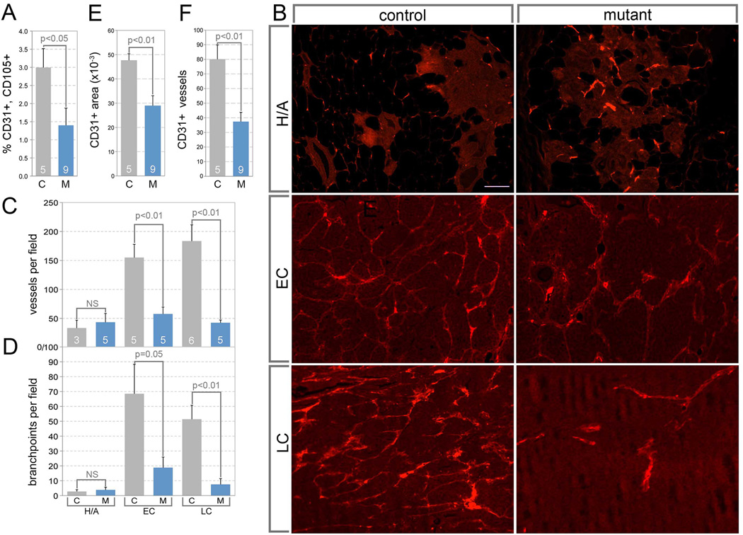
Fig 3. The angiogenic switch is suppressed in the absence of TAM Wnt7b.
(A) Percentage of CD31+, CD105+ vascular endothelial cells in control and mutant tumors combined from the 20–22 week range. (B) Representative images of Texas Red dextran perfused blood vessels in PyMT mammary tumors at premalignant (Hyperplasia and Adenoma, H/A), or malignant (early carcinoma, EC, and late carcinoma, LC) stages. The scale bar in (B), H/A, control is 100 µm and applies to all panels. (C, D) Quantification of dextran labeled vessels for a given tumor stage using either vessels per field (C) or branch-points per field (D). (E, F) Quantification of CD31 labeling in control (C) and mutant (M) late carcinomas shown either as CD31+ area (E) or CD31+ vessels (F). For (A and C–F) sample number is shown at base of histogram bar. Error bars are SEM.

