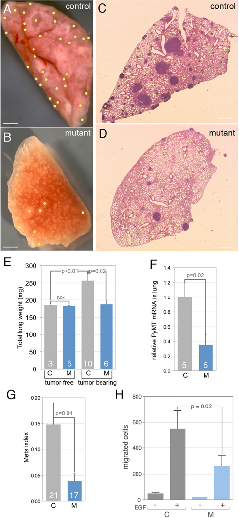
Fig 6. Metastasis is reduced in the absence of tumor stroma cell WNT7b.
(A–D) Representative images of metastasized lung in control (A, C) and mutant (B, D) mice in whole mount (A, B) and in section (C, D) at 22 weeks. Scale bars in (A–D) are 180 µm. (A, B) Obvious surface metastases in whole mount lungs are marked with yellow dots. (C, D) In lung sections, metastases appear as dense purple regions. (E) Quantification of total lung weight in control and mutant, tumor free and tumor bearing mice at 22 weeks. (F) Relative PyMT mRNA expression in control and mutant lung at 22 weeks. (G) The lung metastasis index for control and mutant mice at 22 weeks. Sample number is shown at the base of each histogram bar in (E–G). (H) Quantification of the number of cells that migrate into micro-needles placed in either control (MMTV-PyMT; Wnt7btm2Amc/−, C) or mutant (MMTV-PyMT; Wnt7btm2Amc/−; Csf1r-iCre, M) tumors. The micro-needles were loaded with either vehicle (−) or EGF (+) as labeled. These data show that the absence of TAM Wnt7b significantly reduced the number of cells that migrate into the needle under the influence of EGF. Error bars for all charts are SEM.

