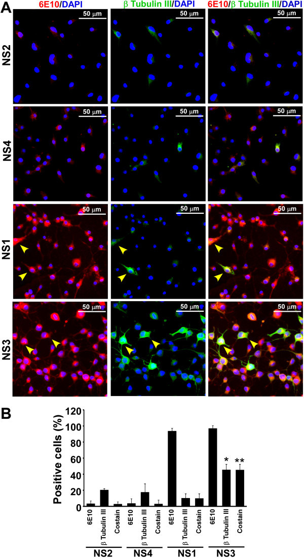
Figure 5.

Expression of APP and β-tubulin III proteins in neurosphere monolayer cultures. (A) Triturated cells from NS cultures were grown on PDL coated optical cover-glass plates or glass coverslips for 3 days in complete media. Immunostaining with 6E10 (specific for human APP) and β-tubulin III antibodies, image acquisition, analysis and display are similar to Figure 2. Images demonstrate presence of cells positive for both APP and β-tubulin III in NS1 and NS3 (yellow arrowheads). (B) Percent cells positive either for APP or β-tubulin III or both (co-stain) are plotted as histograms of mean + standard deviation from three independent data sets. Significantly more APP and β-tubulin III co-stained cells are observed in NS3 line. *indicates p ≤ 0.05 when compared with other neurospheres and **indicates p ≤ 0.05 when compared with NS1 co-stained cells.
