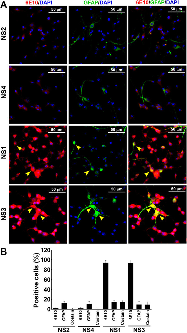
Figure 6.

Expression of APP and GFAP proteins in neurosphere monolayer cultures. (A) Immunostaining of APP and GFAP on cell monolayers were performed as described earlier. Images demonstrate that some cells are positive for both APP and GFAP (yellow arrowheads) in Tg+ve NS lines. (B) Percent cells positive either for APP, GFAP or both (co-stain) are plotted as histograms of mean + standard deviation from four independent experiments.
