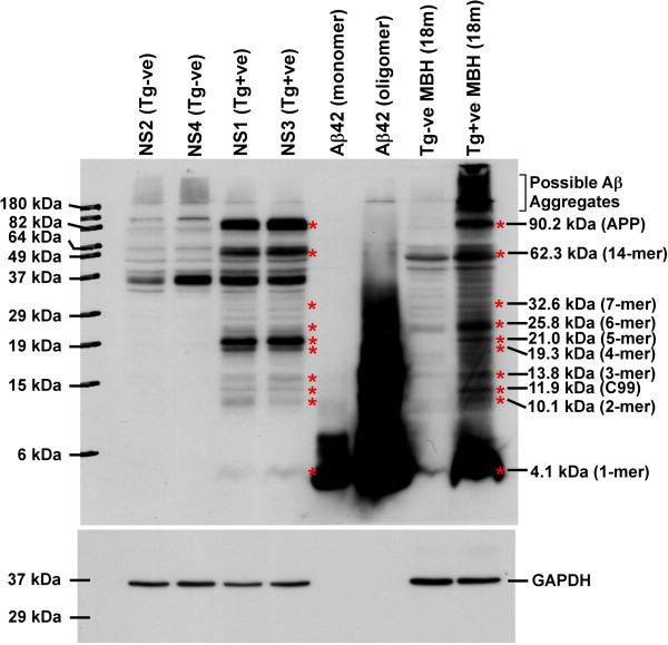
Figure 7.

Expression of huAPP and its proteolytic fragments in Tg+ve neurosphere lines. Sixty microgram of total protein from each NS lysate was western blotted onto 0.2 μm PVDF membrane and immunoblotted with 6E10 antibody after HIER. Images clearly indicate the presence of huAPPswe full-length protein only in NS1, NS3 and 18 month (18 m) old Tg+ve mouse brain homogenate (MBH) but not in NS2, NS4 and 18 m old Tg-ve MBH. Monomeric and 2 days old oligomeric Aβ42 peptides were taken as positive controls. *indicates bands in common between Tg+ve neurosphere lysates and 18 m old Tg+ve MBH but not present in Tg-ve neurosphere lysates and 18 m old Tg-ve MBH. Molecular weight analysis indicates these bands could represent Aβ monomers to various oligomers as mentioned. Membranes were stripped and immunoblotted with GAPDH antibody to verify differences in protein loading. Data represents four independent experiments.
