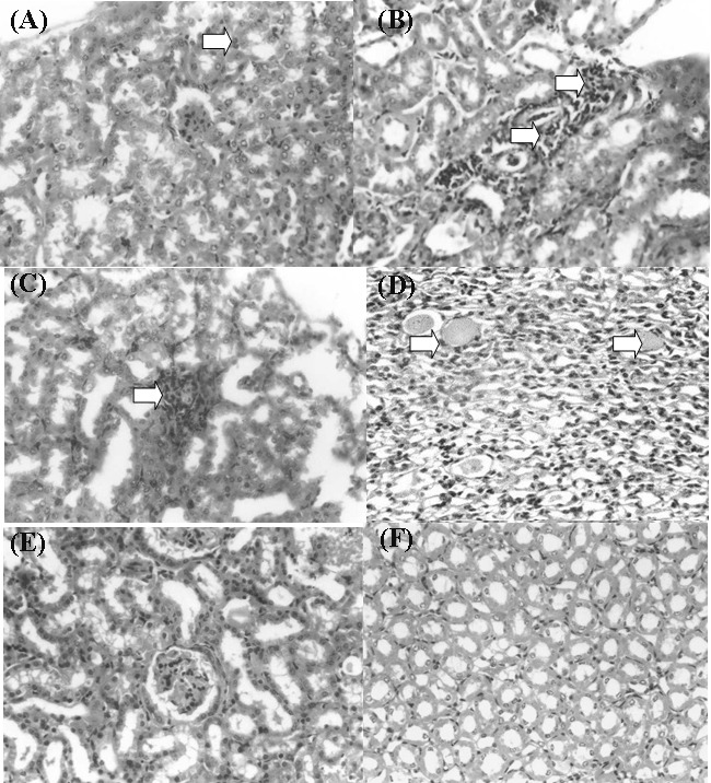
Figure 3 .

Histopathological changes in the kidneys of mice treated with mefenamic acid.
A: Mild interstitial nephritis in the renal cortex of mouse (Arrow) received 200 mg/kg mefenamic acid for 1 day (Mag. 400x)
B: Interstitial nephritis and glomerular necrosis (Arrows) in the renal cortex of mouse received 100 mg/kg mefanamic acid for 14 days (Mag. 400x)
C: Interstitial nephritis in the renal cortex of mouse (Arrow) received 100 mg/kg mefanamic acid for 14 days (Mag. 400x)
D: Medullary tubular atrophy (Arrows) of mouse received 100 mg/kg mefanamic acid for 14 days (Mag. 400x)
E: Section from control mouse showing normal renal cortex histology (Mag. 400x)
F: Section from control mouse showing normal renal medulla histology (Mag. 400x)
