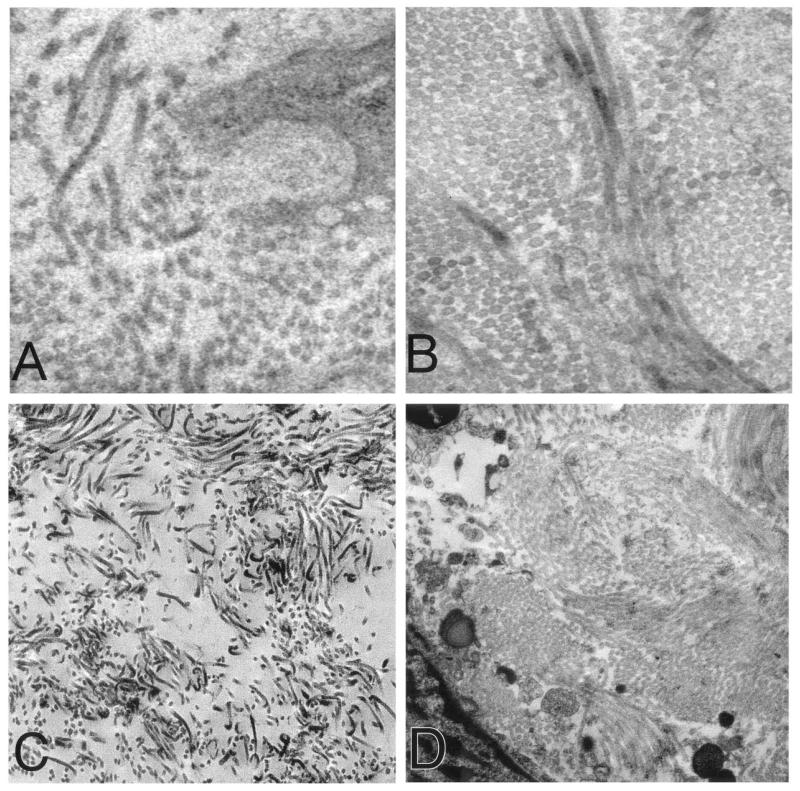
FIGURE 1.
(A), Electron micrograph (×31,000) of leiomyomas demonstrating short, randomly aligned, and widely dispersed collagen fibrils. Tissue sample was obtained from a region near the capsule of the leiomyoma. The subserosal midcavity leiomyoma measured 0.3 × 0.3 × 0.3 cm. This 58-year-old subject had a total of six leiomyomas. She received no hormonal therapy. (B), Electron micrograph (×31,000) of paired myometrium. Collagen fibrils of the adjacent myometrium were closely packed and well aligned in a parallel manner. The myometrium was obtained 0.5 cm from the edge of the leiomyoma. (C), Leiomyoma sample from subject 2 also showing loosely packed collagen fibrils and a random orientation (×12,500). This sample was subserosal and measured 3.8 × 3.5 × 2.8 cm. This 46-year-old subject had a total of four leiomyomas and did not receive hormonal therapy. (D), Electron micrograph (×12,500) of myometrium adjacent to the leiomyoma in C revealing well-packed collagen fibrils.

