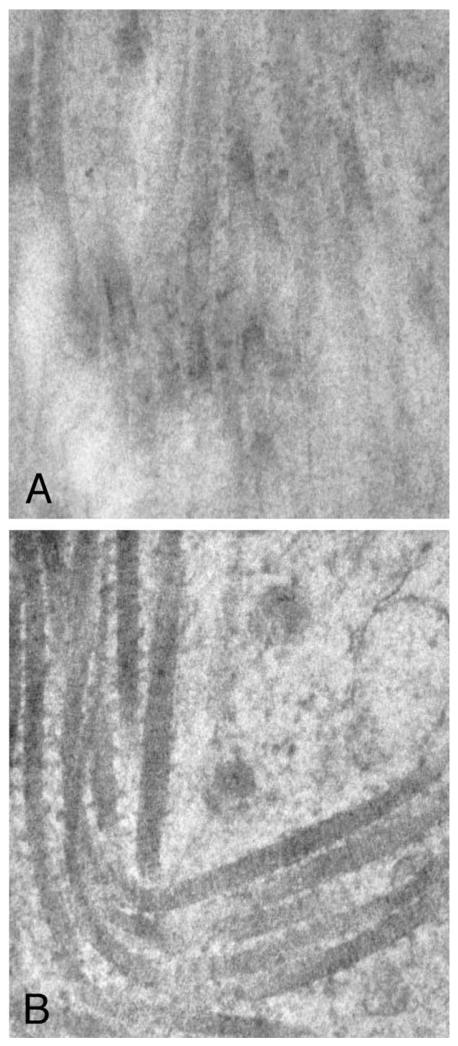
FIGURE 2.
A, Electron micrograph of a leiomyoma (×64,000). Note the disorganization of the collagen fibrils. There was no observed evidence of a barbed appearance of fibrils in the leiomyoma tissue studied. This sample was taken from a 0.3 × 0.3 × 0.3 cm fibroid. Subject 4 was 35 years old and had three leiomyomas. B, At the same magnification, the collagen fibrils in the myometrium have a clear and distinct barbed appearance, suggesting heterotypic fibrils. The myometrium was obtained 2.5 cm from the edge of the leiomyoma. The subject had been taking FeSO4 for uterine bleeding.

