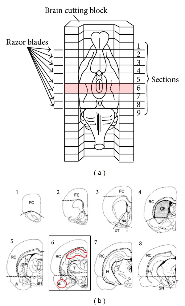
Figure 1.

(a) Orientation of brain within brain cutting block; (b) coronal brain sections from dissected regions. (a) Illustration of rat brain in cutting block depicting specific areas where coronal slices were performed; (b) numbers correspond to dissected sections from (b). FC, frontal cortex; NA, nucleus accumbens; OT, olfactory tubercle; S, septum; CP, caudate putamen; RC, remaining cortex; GP, globus pallidus; aH, anterior hypothalamus; pH, posterior hypothalamus; A, amygdala; T, thalamus; SN, substantia nigra; VT, ventral tegmentum; H, hippocampus [14].
