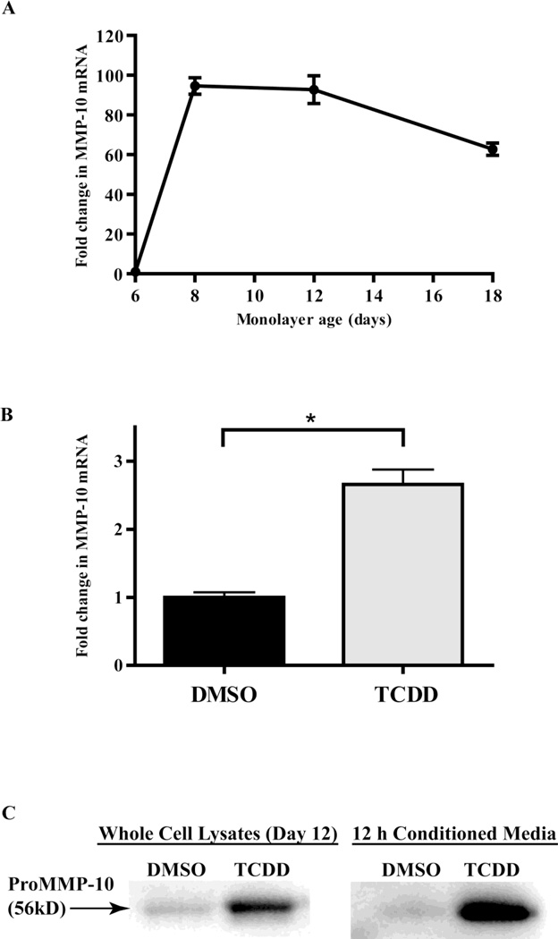
Figure 3. TCDD increases expression of MMP-10 in monolayer cultures of keratinocytes.
(A) NIKS cells were grown without feeder layers in 6 well plates and treated with DMSO. The relative expression level of MMP-10 mRNA was determined by Q-PCR at day 6, 8, 12 and 18 post-treatment. Values were normalized to cyclophilin-A, then expressed as fold induction of the level found in day 6 DMSO treated cells. Data shown are means ± SEM from a single experiment performed in triplicate and are representative of at least 3 independent experiments. (B) The relative expression level of MMP-10 mRNA was calculated by Q-PCR for NIKS treated with DMSO control, or 10nM TCDD, at 12 days post-treatment. The levels of MMP-10, were normalized to cyclophilin-A, then expressed as fold induction over day 6 DMSO treated cells. Data shown are means ± SEM from a single experiment performed in triplicate and are representative of at least 3 independent experiments. *P<0.05 by t-test (C) Whole cell lysates of NIKS treated with DMSO or 10 nM TCDD for 12 days (left panel) or 12 hr conditioned media from these cultures (right panel) were resolved by SDS-polyacrylamide gel electrophoresis and probed using a monoclonal antibody against human recombinant MMP-10 (R&D systems, Minneapolis, MN).

