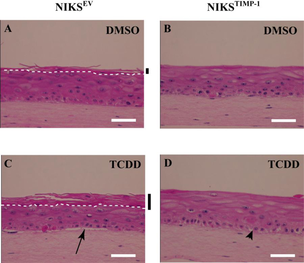
Figure 5. TIMP-1 overexpressing keratinocytes show no change in tissue architecture following TCDD treatment in organotypic culture.
NIKSEV (A,C) and NIKSTIMP-1 (B,D) cells were grown in organotypic culture in the presence of DMSO (A,B) or 10nM TCDD (C,D) for 10 days. Cultures were then fixed, paraffin embedded, sectioned and stained with hematoxylin and eosin. White dashed lines designate the boundary between the stratum corneum and stratum granulosum. Black bars indicate the cornified layer. The arrow highlights separation of the dermis from basal layer. The arrow head designates intercellular spaces. White scale bars each equal 50 µm.

