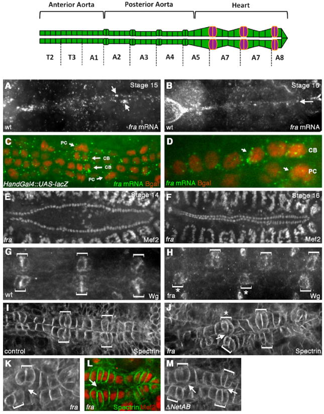
Fig. 1.

fra is expressed in the dorsal vessel and fra mutants have defects in CB alignment and attachment. Top panel shows a schematic of the embryonic DV, which spans segments T2–A8 and is divided into the anterior aorta, posterior aorta and the heart. In the heart, the six pairs of Wingless-expressing CB ostial cells are indicated in magenta. (A, B) In situ hybridization was performed with a DIG-labeled fra antisense probe and visualized using Tyramide signal amplification. Expression of fra at stage 15 is highly enriched in the DV (A). fra expression persists through stage 16 (B). (C, D) Co-staining Hand-Gal4/UAS-lacZ embryos for fra mRNA (green) and β-gal (red) shows the expression of fra mRNA on both the CBs and PCs (arrows). (E-F) anti-DMef2 staining in fra3 mutants at stage 14 (E) and 16 (F) showing that CB specification and migration to the dorsal midline is not impaired in fra3 mutants. Wild type embryos have 104 CBs, and we found no significant difference in CB number in fra3 mutants (104+/−2, n=5). (G-H) Staining with anti-Wingless (Wg) in wild type (wt) (G) or fra3 mutant (H) reveals that the ostial cells (brackets) are correctly specified in fra3 mutants, but are often misaligned across the dorsal midline (asterisks). (I-J) anti-αSpectrin staining in the heart region of the DV. In wild type stage 16 embryos, the ostial cells (brackets) are aligned with each other and make contact across the dorsal midline (I). In fra3 mutants (J), ostial cells (brackets) are often mis-aligned (asterisk) and fail to make proper contact with their contralateral counterparts (arrows). (K) Another fra3 mutant embryo showing mis-aligned CB ostial cells (brackets) and failure of CBs to make contact across the dorsal midline (arrow). (L) fra3 embryo double stained with anti-DMef2 (red) and anti-αSpectrin (green) to show that the space between CBs is not due to the presence of an extra CB. (M) ΔNetAB mutant embryos also show defects in CB attachment (arrows).
