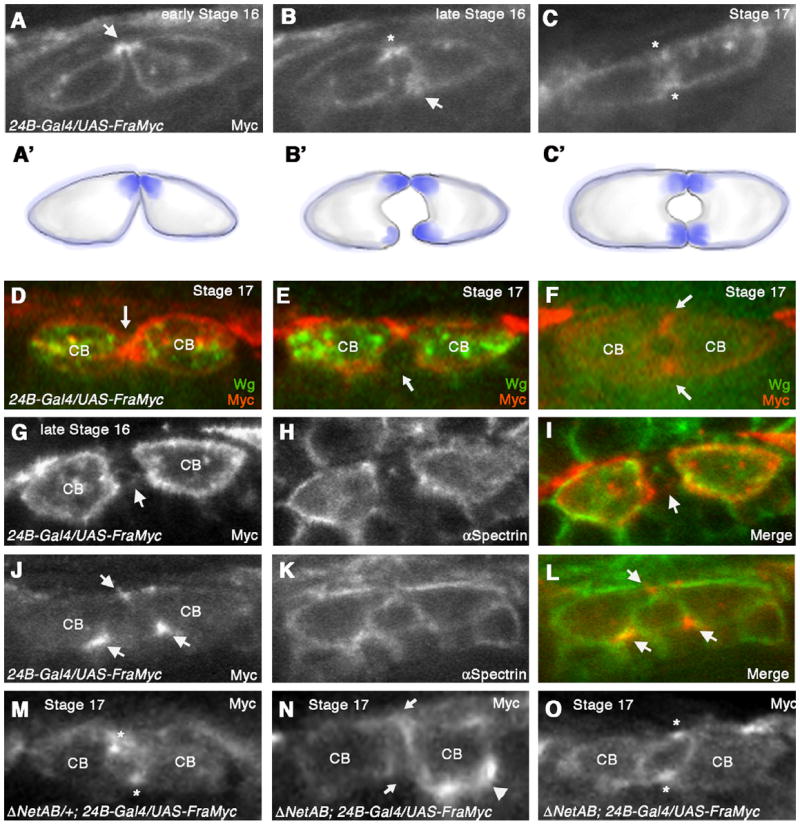
Fig. 4.

Frazzled protein concentrates at sites of CB attachment (A–C) Cross-sections of embryos expressing UAS-fra-Myc in all CBs with 24B-Gal4. (A) Anti-Myc staining at early stage 16 reveals that Fra-Myc accumulates dorsally in the CBs corresponding to the initial sites of contact (arrow). (B) At late stage 16, Fra-Myc also accumulates at the site of ventral CB outgrowth (arrow). Dorsal contact is indicated with an asterisk. (C) Stage 17 embryo when lumen formation is complete. Fra-Myc staining persists at dorsal and ventral sites of contact (asterisks). Schematic diagrams illustrating Fra-Myc localization (A’–C’). (D, F) Double labeling of UAS-fra-Myc/24B-Gal4 embryo with anti Myc and anti-Wg, which labels the 6 pairs of ostial CBs in the heart. CBs that were Wg positive showed CB attachment phenotypes as well as uniform distribution of Fra-Myc (D and E). In CBs that were negative for Wg protein (F), Fra-Myc accumulated at dorsal and ventral points of attachment (arrows) and the lumen appeared normal. (G-I) UAS-fra-myc/24B-Gal4 embryo sectioned through the ostial CBs, which express high levels of Fra-Myc. In these embryos, Fra-Myc staining (G) was evenly distributed along the entire CB membrane, similar to αSpectrin (H). (I) is a merge of (G) and (H). (J–L) UAS-fra-myc/24B-Gal4 embryo sectioned through the non-ostial CBs expressing lower levels of fra-Myc. Arrows point to the areas that accumulate Fra-Myc protein. (L) is a merge of (J) and (K). (M–O) (M) Fra-Myc accumulates at dorsal and ventral attachment points (asterisks) in a ΔNetAB heterozygote at stage 17. (N) In a ΔNetAB mutant, Fra-Myc accumulation at these sites is disrupted (arrows) and we observe inappropriate accumulation of Fra-Myc on areas of the CB membrane not normally associated with attachment (arrowhead). In these sections a proper lumen fails to form. (O) In ΔNetAB mutants that show a normal lumen, we also observe normal distribution of Fra-Myc (asterisks).
