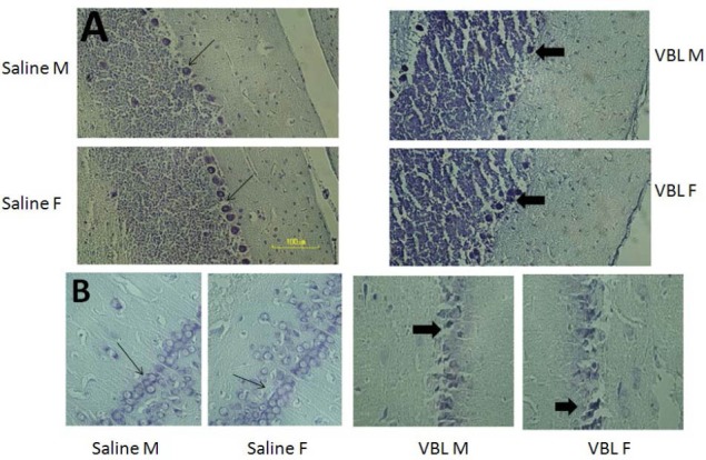
Figure 4.

Neurons from the cerebellar cortex and hippocampus 5 weeks after exposure to Vinblastin (VBL). Most neurons from the cerebellum (A), and the hippocampus (B) in saline-treated rats have normal morphology (arrows), but for the VBL-treated groups, several degenerated cells (arrowheads) can be seen with shrinkaged nuclei and dark cytoplasm
