
Figure 5.
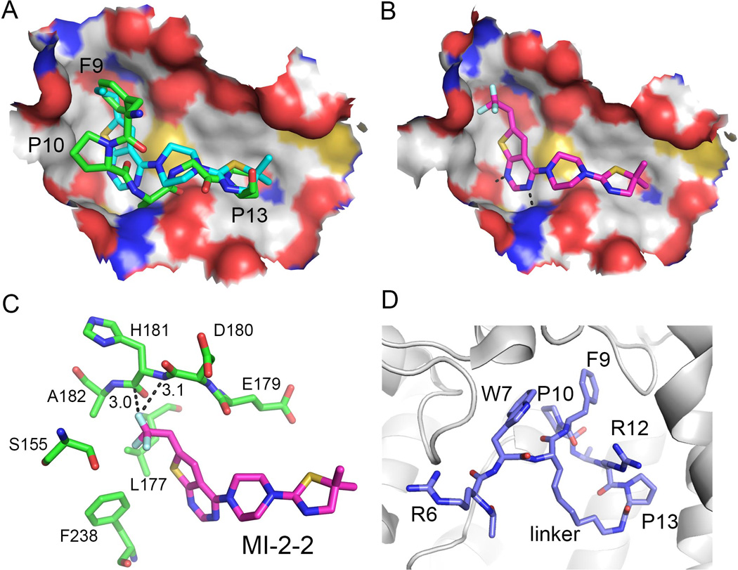
Structures of small molecule inhibitors bound to menin. A. MI-2 (cyan carbon atoms, PDB code 4GQ3) inhibitor mimics key interaction of MBM1 (green carbon atoms, PDB code 4GQ6) with menin. F9, P10 and P13 pockets are labeled. B. Binding mode of MI-2-2 to menin (4GQ4). C. Trifluoroethyl group in MI-2-2 forms favorable interactions with menin backbone; short distances between fluorine atom and backbone are labeled. D. Structure of the macrocyclic MLL peptide (blue carbon atoms, PDB code 4I80) bound to menin (shown as gray ribbon). Position of the linker used for cyclization of residues 8 and 13 is labeled.