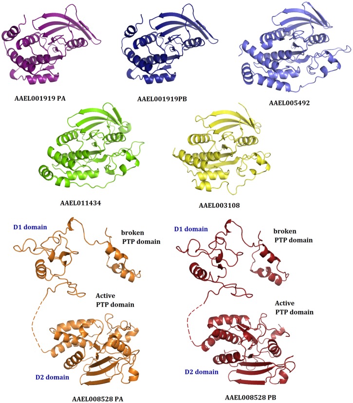
Figure 3. Structural models of the ‘active’ PTP sequences from A. aegypti.
All proteins showed the classical PTP fold with a twisted β sheet at the center surrounded by α helices. Because of its low sequence conservation, the absent PTP domain of AAEL008528 could not be modelled completely. Only one model is shown for genes with splicing variants because their PTP domains are identical. For additional details, please check Figure 4.

