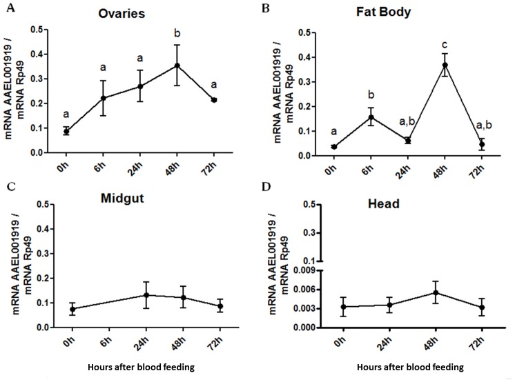
Figure 8. Expression of AAEL001919 after blood feeding.
Five- and seven-day old female mosquitoes (20–30 insects) were naturally fed with rabbit blood and dissected 24 h, 48 h and 72 h later. The following tissues were evaluated for AAEL001919 gene expression: (A) Ovaries; (B) Fat body; (C) Midgut and (D) Head. Non-blood fed mosquitoes (0 h) were maintained on 10% sucrose ad libitum and were concomitantly dissected at any one of the three time points. Normalized data from five experiments were analyzed by one-way analysis of variance (ANOVA) and by a post-test Tukey's Multiple Comparison Test (a, b, c p<0.05). Groups assigned with the same letter (a, b or c) indicate that they do not show statistically significant differences among them.

