
Figure 4.
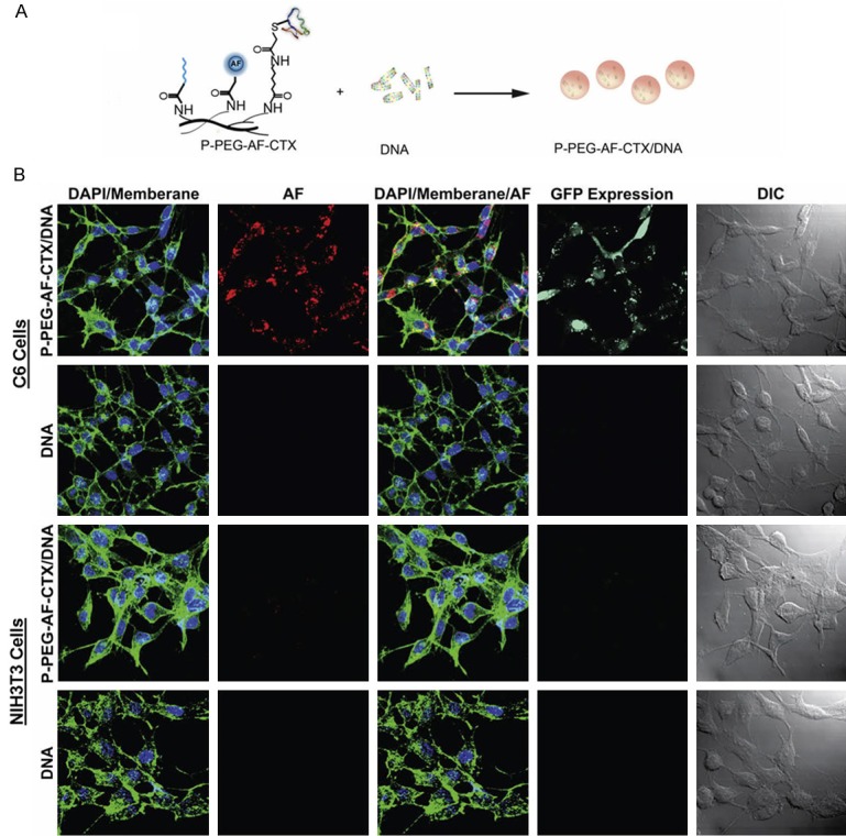
A: The polymeric construct complexed with DNA to generate the targeting nanovector (P-PEG-AF-CTX/DNA). B: Confocal fluorescence and differential interference contrast (DIC) images of C6 and NIH3T3 cells treated with 10 µg DNA mL-1 without a delivery vector (DNA) or with vectors complexed with PEGylated and CTX-enabled PEI (P-PEG-AF-CTX/DNA). Cellular membranes are shown in green, nuclei in blue, polymeric vectors in red, and GFP expression in turquoise. Scare bars correspond to 40 µm. Reprinted with the permission of the Journal of Biomaterials, Veiseh et al., 2009.