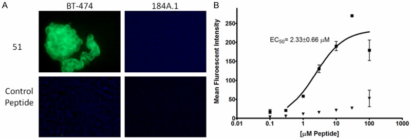
Figure 2.

Peptide 51 In Vitro Cell Binding Assays. A: Biotinylated peptide 51 and a control peptide were incubated with BT-474 human breast cancer and 184A.1 normal breast epithelial cells fixed onto microscope slides. Following washing, bound peptides were detected by addition of an anti-biotin Alexafluor 488-conjugated antibody. Strong binding is observed for 51 with the target BT-474 cells, but not normal breast epithelial cells. The control peptide does not exhibit binding to either cell line. B: Peptide 51 was analyzed for BT-474 and 184A.1 specificity and affinity by flow cytometry. Peptides were diluted from 100 nM to 100 µM in 0.1% TBST. Following incubation of peptide with cells, bound peptide was detected by anti-biotin Alexafluor 488. Squares represent the mean of 3 BT-474 replicates at the indicated peptide concentration, triangles represent the mean of 3 184A.1 replicates. Error bars represent the standard deviation.
