
Figure 2.
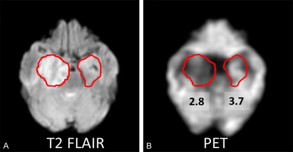
A: A 60-year-old male patient (case 7) shows abnormal signal and enlargement in right medial temporal lobe on T2 FLAIR image, as compared to the contralateral ROI. B: The co-registered FDG-PET and MRI image shows that this lesion has lower SUV (SUV=2.8) than contralateral homologous cortical region (SUV=3.7).