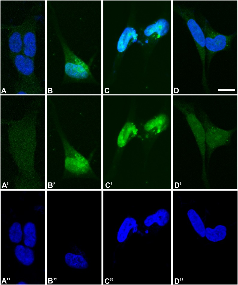
Figure 3. Indirect immunofluorescence to detect HB9 protein in SK-N-BE cells.
A, B, C, and D show SK-N-BE cells at proliferative stage (day 0) and at 4th day, 5th day, and 6th day after acid retinoic induction respectively. HB9 protein (green signal) is visible in both nucleus and cytoplasm at the fourth and fifth days of differentiation (B, C). A very faint staining is also noted in the cytoplasm of cells at proliferating stage (A) and in the cytoplasm of cells at the sixth day of differentiation (D). A, B, C and D are the merge images of A’, B’, C’, D’, representing immunofluorescence staining only, and A”, B”, C”, D”, representing the DAPI staining of the nucleus only in blue. Scale bar is 10 µm.

