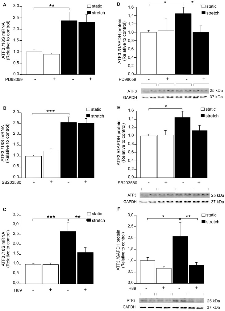
Figure 5. The effect of kinase inhibitors on mechanical stretch–induced increase in ATF3 expression.
ERK inhibitor PD98059 (A, D), p38 inhibitor SB203580 (B, E) or PKA inhibitor H89 (C, F) were added at the concentration of 10 µM each. DMSO was used as a control. Approximately 2 hours after the insertion of inhibitors or DMSO, the cell cultures were subjected to cyclic mechanical stretching for 1 hour. ATF3 mRNA levels were determined by RT-qPCR and normalized to 18S quantified from the same samples (A–C). The mRNA levels are presented relative to non-stretched DMSO control cells. The results represent mean ± SEM (n = 2–19) from at least 3 independent experiments. The expression levels of ATF3 protein and GAPDH loading control were detected by Western blotting (D–F). Representative Western blots are shown. ATF3 protein levels were normalized with GAPDH levels and are presented relative to non-stretched DMSO control cells. Bar graphs represent mean ± SEM (n = 4–12) from at least 3 independent experiments. *P<0.05; **P<0.01; ***P<0.001.

