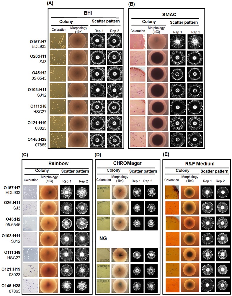
Figure 2. Representative images of colony coloration, microscopic colony morphology and scattering patterns of E. coli strains grown on each of the following medium: (A) BHI agar, (B) Sorbitol MacConkey agar (SMAC), (C) Rainbow, (D) CHROMagar O157, (E) R&F medium.
All colony scatter images were captured at 10–12 h of incubation when colonies reached optimum size. *NG: no growth of O103 strains on CHROMagar.

