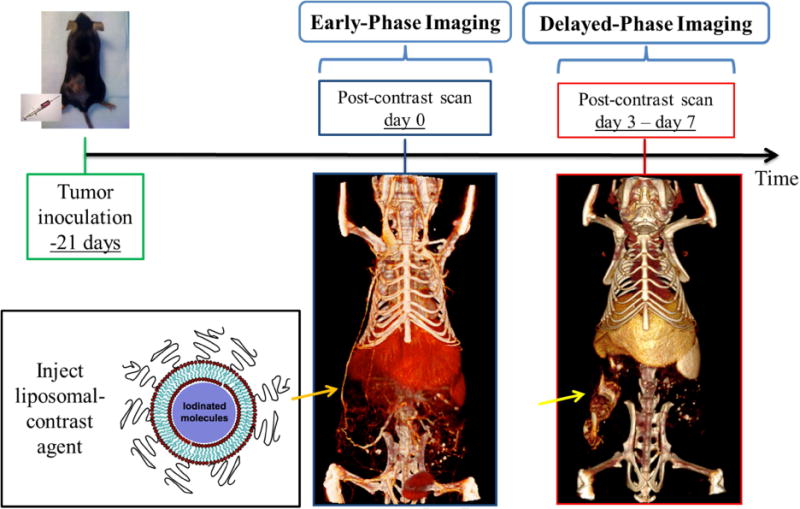
Fig. 5.

In vivo cancer imaging using liposomal iodinated nanoparticles. Mice are inoculated with cancer cells 3 weeks prior to imaging, allowing xenograft tumors to grow. Imaging is performed immediately after the injection of liposomal iodinated nanoparticles (early phase) and 3 to 7 days later (delayed phase). The early-phase scan is used to measure tumor fractional blood volume, while the delayed-phase scan is used to measure tumor vessel permeability via the enhanced permeability and retention effect.
