
Fig. 8.
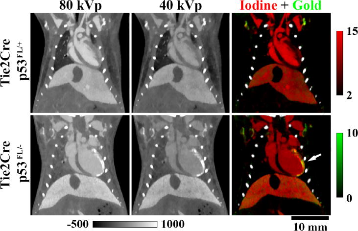
In vivo, dual energy micro-CT used to assess accumulation of gold nanoparticles in the left ventricle of mice after partial heart irradiation. Iodine maps (red) were used to contour the total volume of the left ventricle, whereas gold maps (green) from the same animal were used to quantify the volume of the myocardium that had increased gold nanoparticle accumulation (white arrow). Concentrations of iodine and gold are in mg/ml. Grayscale images are scaled in Hounsfield units. The genotypic backgrounds (i.e. Tie2Cre p53FL/− and p53FL/+) are detailed in [165].