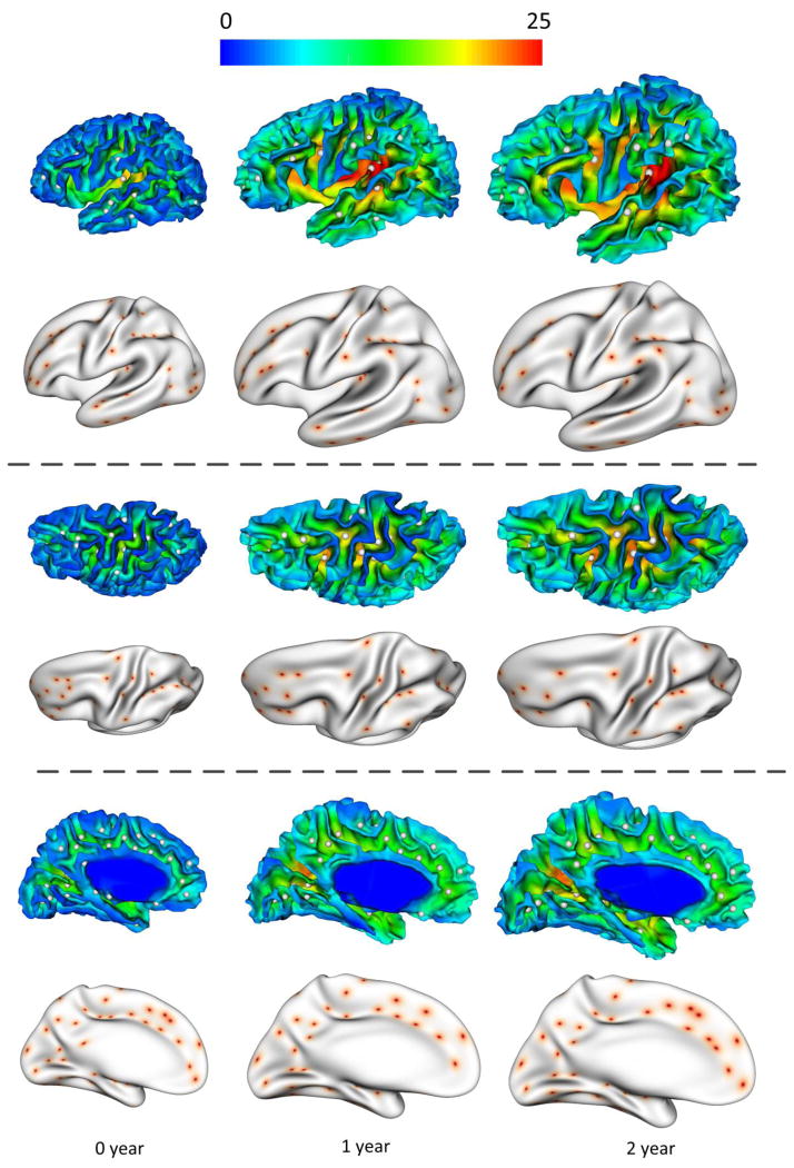
Figure 2.
Sulcal pits extraction results on the left hemisphere of a representative infant at 0, 1 and 2 years of age. In the first, third and fifth rows, the sulcal pits represented by white points are overlaid on the cortical surfaces, color-coded by the sulcal depth (mm). For better inspection, in the second, fourth and sixth rows, the sulcal pits represented by red points are mapped onto the partially inflated cortical surfaces.

