
Figure 1.
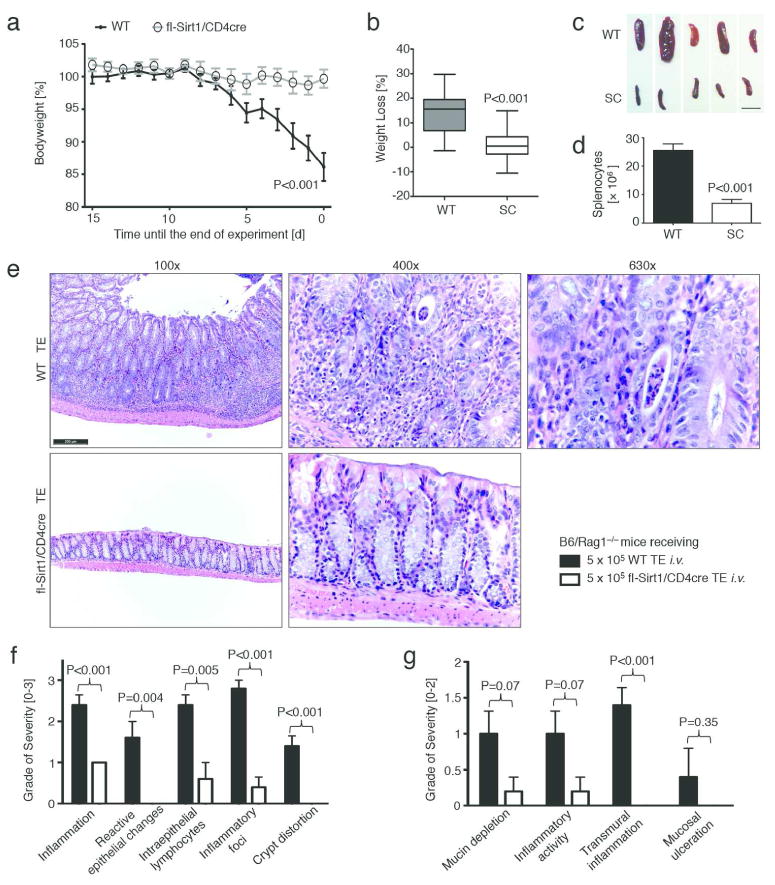
T effector cells lacking Sirt1 cause less autoimmune colitis. Twelve weeks old B6/Rag1−/− mice were adoptively transferred with 5 × 10⁵ CD4+CD25− (<1.1% Foxp3+) T effector cells (TE) i.v. of either C57Bl/6 wild type (WT) or fl-Sirt1/CD4cre (SC) origin and followed until autoimmune colitis developed. Data pooled from four independent experimental setups (n=34 mice). (a, b) B6/Rag1−/− mice injected with SC TE developed less disease and weight loss. (a) Weight curves, normalized to the end point of each of the three independent experiments. (b) Percent weight loss as boxplots (whiskers: 5th-95th percentile). (c) Smaller spleen size in fl-Sirt1/CD4cre TE injected B6/Rag1−/− mice. Scale bar indicates 1 cm. (d) B6/Rag1−/− mice receiving WT TE showed a higher spleenocyte count than fl-Sirt1/CD4cre TE recipients. (e) Colonic specimens from B6/Rag1−/− mice receiving WT TE show significant colitis including marked crypt architectural distortion (100x original magnification) with neutrophilic infiltration of the lamina propria, crypt epithelium (“cryptitis”) and glandular lumina (“crypt abscess”, see 630x original magnification). In contrast, adoptive transfer of TE lacking Sirt1 produced much less signs of colitis. H&E staining; scale bar indicates 200 μm. (f, g) Pooled data from blinded histologic analysis demonstrate less colitis pathology in mice adoptively transferred with fl-Sirt1/CD4cre TE (n=10).