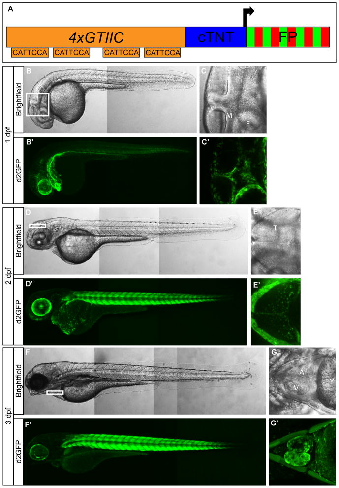
Figure 1. 4xGTIIC:d2GFP expression during zebrafish development.
(A) Schematic of the Yap/Taz-Tead reporter construct. (B′,D′,F′) Low magnification images show 4xGTIIC:d2GFP expression in whole embryos/larvae at 1 dpf (B′), 2 dpf (D′) and 3 dpf (F′). (C′, E′, G′) Higher magnification images show d2GFP expression in non-neural cells located at the (C′) midbrain-hindbrain boundary and around the eye at 1 dpf, within the tectum at 2 dpf (E′), and in the heart at 3 dpf (G′). Brightfield images (B,C,D,E,F,G) are placed above each fluorescent image. cTNT=chicken troponin T minimal promoter; FP= fluorescent protein; M=midbrain; H=hindbrain; E=eye; A=Atrium; V=Ventricle; Y=Yolk. B, B′, C, C′, D, D′, F, F′ = lateral view; E, E′ = dorsal view; G, G′ = ventral view.

