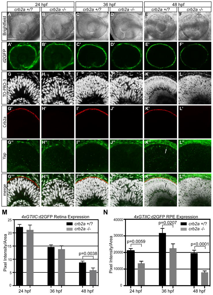
Figure 6. Loss of crb2a results in a decrease of 4xGTIIC:d2GFP expression in the neural retina at 48 hpf and at 24, 36, 48 hpf in the RPE.
(A–F′) Neural retina and RPE 4xGTIIC:d2GFP expression in crb2a +/? and crb2a −/− embryos. (G–L‴) Immunofluorescence of Yap (green) and Crb2a (red) in crb2a +/? and crb2a −/− embryos. Arrow indicates Yap positive cells in the crb2a +/? neural retina that do not appear in crb2a −/−. (M,N) Quantification of d2GFP pixel intensity in the (M) neural retina and (N) RPE of crb2a +/?/crb2a −/− embryos at 24 (n=46/n=13), 36 (n=32/n=28), and 48 (n=26/n=23) hpf. Error bars represent S.E.M. p=unpaired t-test with equal S.D. Scale bar=50μm in A,C, and E.

