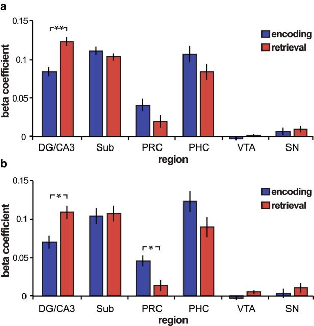
Figure 4.
Task-related changes in background connectivity. a, Area CA1 background connectivity during the Encoding Task (blue) and Retrieval Task (red). Hippocampal areas DG/CA3 and CA1 had significantly greater functional connectivity during Retrieval Task blocks than during Encoding Task blocks (p = 0.003). b, CA1 functional connectivity adjusting for task order. Connectivity was re-estimated dropping the first Encoding Task run and the last Retrieval Task run to adjust for task order. Significant task differences were found for CA1–DG/CA3 functional connectivity (t(15) = 2.3; p = 0.04) and for CA1-perirhinal (PRC) functional connectivity (t(15) = 2.27; p = 0.04). Error bars indicate the SE of the difference score. *p < 0.05; **p < 0.005. SN, Substantia nigra; PHC, parahippocampal cortex.

