
Figure 6.
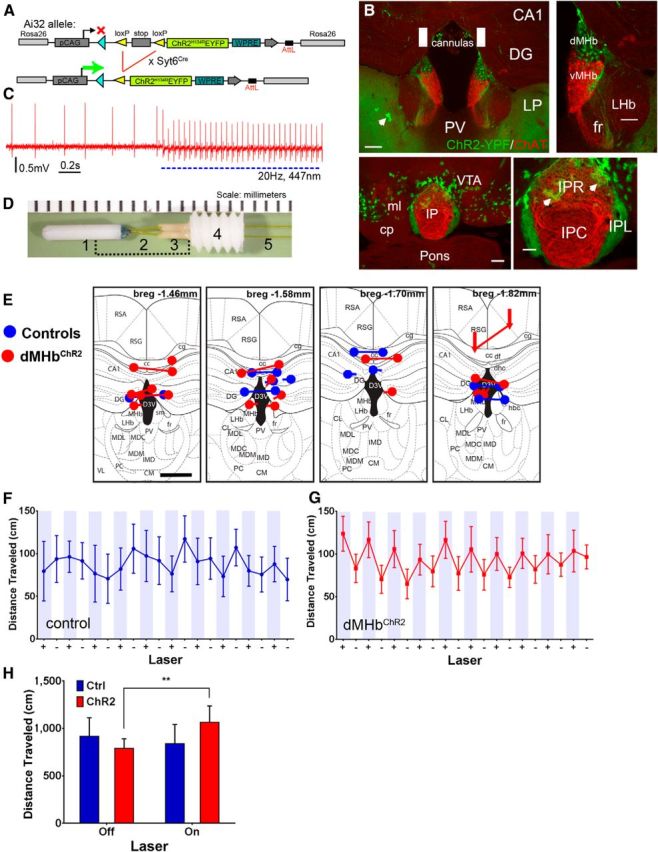
An optogenetic model for dMHb function and the effect of dMHb stimulation on locomotion. A, Transgenic strategy for conditional expression of ChR2-EYFP in the dMHb. B, Syt6Cre-driven expression of ChR2-EYFP from the Ai32 reporter in the dMHb (top) and IPL (bottom). EYFP expression is also seen in some scattered cells, which have the appearance of astrocytes (arrows). EYFP-labeled terminal fibers are also observed in the lateral posterior thalamic nucleus (LP); these are projections from layer 5/6 cortical neurons. No EYFP-labeled fibers or cell bodies are observed in the paraventricular thalamus immediately ventral to the IP. No labeled fibers are observed in the VTA or other tegmental areas adjacent to the IP. White rectangles represent the targeted position and size of the implanted fiber optic cannula. Scale bar: low-power views, 200 μm; high-power views, 100 μm. C, Loose-seal cell-attached recording showing light-induced action potentials in the dMHb in a dMHbChR2 mouse. Irregular spontaneous firing at ∼3 Hz is observed at baseline. Application of 447 nm blue light pulses (20 ms, 2.0 mW/mm2) at 20 Hz elicits action potentials entrained to the pulse frequency. The maximum stimulation rate for which the cell shown would generate 1:1 action potentials was ∼20 Hz; at higher stimulation frequencies, some light pulses failed to elicit spikes. At the end of a 10 s interval of pulsed light delivery, a period of suppressed firing was observed (∼7 s, data not shown), followed by a gradual resumption of firing at the baseline rate. D, Bilateral optical cannula for implantation in the dMHb. Cannula consists of a zirconia ferrule (1) and a 0.1 mm polyimide-coated optical fiber (2,5), a polyether ether ketone (PEEK) insert (3), and an acetal (Delrin) guide (4). The bracketed area is covered with a stainless steel protective sleeve before implantation. E, Optical fiber placement in the habenula for in vivo optogenetic-stimulated control and dMHbChR2 mice. Blue represents fiber placement in control mice; red represents placement in dMHbChR2 mice. Fiber termini are shown on the level of a standard anatomical map (Paxinos and Franklin, 2001) closest to their rostrocaudal position at bregma −1.46, 1.58, 1.70, or 1.82 mm. Nearly all of the cannulas thus were positioned within ±0.2 mm of the intended coordinates at bregma −1.6 mm. Connected dots indicate the probable ventral termini of the optical fibers from each case. In some cases, the right and left optical fibers mapped most accurately to different planes of section and are shown by disconnected dots. If the cannula track could not be followed for the entire length of the optical fiber, the most ventral position and the direction of the cannula track observed are indicated by an arrow. In all cases, the optical fibers were intact and transmitted light efficiently when examined postmortem after the experimental protocol. Scale bar, 0.5 mm. F, G, Distance traveled by control and dMHbChR2 mice during a 10 min open field trial with intermittent laser stimulation. + (shaded), 30 s laser-on periods; −, 30 s laser-off periods. Mice were exposed to the open field and to laser stimulation before testing began to minimize effects of novelty. H, Summary of open field locomotor data. The distance traveled is summed across all laser-on and laser-off epochs for each mouse. In a within-subjects comparison, the dMHbChR2 mice show significantly greater distance traveled during the laser-on than the laser-off periods, whereas control mice exhibit no difference. **p < 0.01, significant difference between laser on/off periods for dMHbChR2 mice. N = 7 control and N = 11 dMHbChR2 mice. cp, cerebral peduncle, basal part; DG, Dentate gyrus; fr, fasciculus retroflexus; IPC, interpeduncular nucleus, caudal; IPL, interpeduncular nucleus, lateral; IPR, interpeduncular nucleus, rostral; LHb, lateral habenula; LP, lateral posterior nucleus of thalamus; ml, medial lemniscus; PV, paraventricular nucleus of thalamus; VTA, ventral tegmental area.